
When a healthcare facility needs clear diagnostic images, the question is not simply whether CT or MRI produces the “better” image. What’s the difference between a CT scan and an MRI for image quality and detail? The answer depends on what needs to be examined, how quickly imaging is needed, the type of tissue involved, and how well the imaging system is maintained.
CT and MRI use different technologies and each has its own strengths. CT is known for fast imaging and strong detail of bones and many internal structures. MRI is especially valuable for showing differences between soft tissues. Both depend on properly working equipment, suitable imaging protocols, and ongoing quality control.
For healthcare facilities, understanding these differences is useful not only when selecting an imaging method, but also when managing the equipment that produces these images.
What’s the Difference Between a CT Scan and an MRI for Image Quality and Detail?
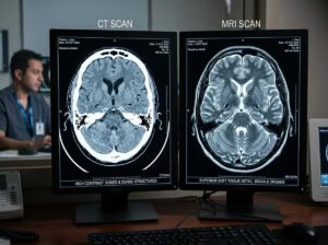
CT and MRI create images in different ways, so neither is always better than the other. CT uses X-rays and computer processing to create detailed cross-sectional images, while MRI uses a powerful magnetic field, radiofrequency energy, and a computer to produce detailed images of the body. MRI does not use ionizing radiation, while CT does. The most appropriate imaging method depends on the body area being examined, the clinical question, and the information needed for diagnosis (RadiologyInfo.org, 2026).
CT is often useful when speed and detailed views of structures such as bone are important. MRI generally provides stronger soft-tissue contrast, making it valuable for areas such as the brain, spine, joints, muscles, and other soft tissues.
How CT Produces Detailed Images
A CT scanner sends X-rays through the body from different angles. Detectors collect the information, and a computer processes it into cross-sectional images.
This allows clinicians to examine structures from different views without relying on a single flat image. CT can be particularly useful for trauma, bone injuries, lung imaging, and situations where fast imaging is important.
However, image quality is not determined by the scanner alone. Patient movement, scanning protocols, reconstruction settings, detector performance, calibration, and artifacts can all affect the final image.
How MRI Produces Detailed Medical Images
MRI works differently. It uses a powerful magnetic field, radiofrequency energy, and computer processing to create detailed images. Unlike CT, MRI does not use ionizing radiation (RadiologyInfo.org, 2026).
MRI is often valuable when doctors need strong contrast between different types of soft tissue. It can be used for brain and spinal imaging, joint problems, muscles, ligaments, tendons, and many other soft-tissue examinations.
MRI image quality can also be affected by patient movement, magnetic-field performance, RF coils, signal-to-noise ratio, positioning, scanning sequences, and artifacts.
For a healthcare facility, this means that maintaining the MRI system is an important part of supporting consistent imaging performance.
CT vs. MRI Image Quality: What Are the Main Differences?
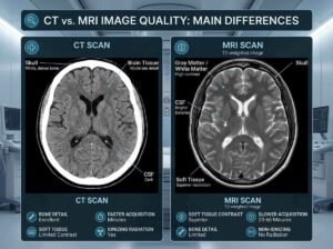
The easiest way to understand CT vs. MRI image quality is to look at where each technology has an advantage.
| Factor | CT | MRI |
| Technology | X-rays | Magnetic field and radiofrequency signals |
| Radiation | Uses ionizing radiation | Does not use ionizing radiation |
| Bone detail | Excellent for many applications | Less commonly the first choice for detailed bone imaging |
| Soft-tissue contrast | Good for many examinations | Generally excellent |
| Speed | Generally faster | Usually takes longer |
| Common strengths | Bone, trauma, lungs, urgent imaging | Brain, joints, spine, muscles, soft tissue |
| Equipment concerns | Tube, detectors, calibration, artifacts | Coils, magnetic-field performance, RF systems, artifacts |
This comparison does not mean that every CT examination is better for bone or every MRI examination is better for soft tissue. Imaging decisions should be based on the patient’s needs and the clinical question.
CT vs. MRI for Bone Detail
CT is often a strong choice when detailed bone anatomy is needed. Its cross-sectional images can show fractures and complex bone structures clearly.
CT vs. MRI for Soft-Tissue Detail
MRI is often preferred when detailed soft-tissue contrast is important. It can help distinguish normal and abnormal tissues in areas such as the brain, spine, and joints.
CT vs. MRI for Speed
CT is generally faster, which can be important in emergency and time-sensitive situations. MRI examinations often take longer and require the patient to remain still for an extended period.
RadiologyInfo.org notes that CT’s fast imaging can be especially helpful for patients who have difficulty remaining still, while MRI produces detailed images without using X-rays or ionizing radiation (RadiologyInfo.org, 2026).
Why Do CT and MRI Images Sometimes Have Poor Quality?

Even a high-end imaging system can produce poor or inconsistent images when something is wrong with the scanning process or equipment.
Patient Movement
Movement can introduce blur or other artifacts. This can be especially challenging during longer MRI examinations.
Imaging Protocols
Image quality also depends on selecting appropriate scanning parameters, positioning the patient correctly, and using suitable reconstruction or imaging sequences.
Equipment Problems
Recurring artifacts or changes in image quality may also point to equipment-related problems. Calibration issues, worn components, detector problems, RF coil issues, or other system faults can affect performance.
This is why healthcare facilities should not automatically treat recurring image-quality problems as normal. They may require further testing or service.
How Quality Control Helps Maintain CT and MRI Image Quality
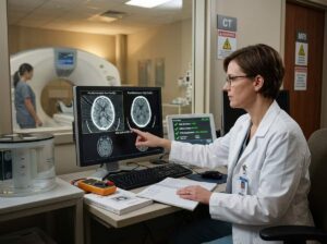
Quality control (QC) is an important part of diagnostic imaging equipment management. It involves regularly checking whether equipment continues to perform within established limits.
For CT, the American College of Radiology (ACR) recommends a documented QC program under the supervision of a qualified medical physicist. Its guidance includes testing areas such as image quality, artifacts, CT number accuracy, spatial resolution, and display calibration.
MRI also requires ongoing QC. ACR guidance includes monitoring areas such as table positioning, image quality, and other system performance measures, with qualified medical physicist or MR scientist involvement.
Quality control and equipment maintenance work together. QC can identify performance changes, while qualified service professionals can help investigate and correct equipment-related problems.
CT and MRI Equipment Maintenance for Consistent Image Quality
Preventive maintenance is more than waiting for a scanner to stop working. Regular service can help healthcare facilities identify problems early, reduce unexpected downtime, and support reliable equipment performance.
For CT systems, ACR guidance states that preventive maintenance should be scheduled, performed, and documented by a qualified service engineer. It also recommends appropriate QC procedures following certain major repairs.
Facilities should pay attention to warning signs such as:
- Recurring image artifacts
- Changes in image quality
- Repeated system errors
- Unexpected equipment behavior
- Failed or out-of-limit QC results
- Repeated component failures
- Unplanned downtime
The goal is not simply to repair equipment after failure. A strong maintenance strategy helps healthcare teams keep critical medical equipment reliable throughout its service life.
Choosing a Biomedical Equipment Service Provider
When selecting a biomedical equipment service provider, healthcare facilities should consider technical experience, preventive maintenance capabilities, repair support, calibration and testing, documentation, and response times.
Syracuse Biomedical Services, LLC provides biomedical equipment maintenance, calibration, repair, preventive maintenance, and on-site and in-house service solutions for healthcare facilities. The company is based in Syracuse, New York, and serves healthcare facilities in Syracuse and surrounding Central New York areas.
Its services are designed to help facilities maintain reliable equipment, reduce downtime, and support ongoing equipment management.
Keep Your CT and MRI Equipment Performing Reliably
CT and MRI systems play an important role in modern diagnostic imaging. Understanding what’s the difference between a CT scan and an MRI helps healthcare professionals recognize which technology is suited to different imaging needs. But producing consistent images also depends on equipment condition, quality control, calibration, and timely maintenance.
If your healthcare facility is experiencing recurring equipment problems, image-quality concerns, or unexpected downtime, professional biomedical equipment support can help identify the issue and keep your systems operating reliably.
Syracuse Biomedical Services offers preventive maintenance, calibration, repair, compliance testing, and biomedical equipment support for healthcare facilities in Syracuse and Central New York.


